Recent advances in Bio-medical

February 25, 2010

Recent developments in biomedical optics

Filed under: Uncategorized — bmeforum @ 5:50 am

The rapid growth in laser and photonic technology has resulted in new tools being proposed and developed for use in the medical and biological sciences. Specifically, a discipline known as biomedical optics has emerged which is providing a broad variety of optical techniques and instruments for diagnostic, therapeutic and basic science applications. New laser sources, detectors and measurement techniques are yielding powerful new methods for the study of diseases on all scales, from single molecules, to specific tissues and whole organs. For example, novel laser microscopes permit spectroscopic and force measurements to be performed on single protein molecules; new optical devices provide information on molecular dynamics and structure to perform `optical biopsy’ non-invasively and almost instantaneously; and optical coherence tomography and diffuse optical tomography allow visualization of specific tissues and organs. Using genetic promoters to derive luciferase expression, bioluminescence methods can generate molecular light switches, which serve as functional indicator lights reporting cellular conditions and responses in living animals. This technique could allow rapid assessment of and response to the effects of anti-tumour drugs, antibiotics, or antiviral drugs.

This issue of Physics in Medicine and Biology highlights recent research in biomedical optics, and is based on invited contributions to the International Conference on Advanced Laser Technology (Focused on Biomedical Optics) held at Cranfield University at Silsoe on 19–23 September 2003. This meeting included sessions devoted to: diffuse optical imaging and spectroscopy; optical coherence tomography and coherent domain techniques; optical sensing and applications in life science; microscopic, spectroscopic and opto-acoustic imaging; therapeutic and diagnostic applications; and laser interaction with organic and inorganic materials.

Twenty-one papers are included in this special issue. The first paper gives an overview on the current status of scanning laser ophthalmoscopy and its role in bioscience and medicine, while the second paper describes the current problems in tissue engineering and the potential role for optical coherence tomography. The following seven papers present and discuss latest developments in infrared spectroscopy and diffuse optical tomography for medical diagnostics. Eight further papers report recent advances in optical coherence tomography, covering new and evolving methods and instrumentation, theoretical and numerical modelling, and its clinical applications. The remaining papers cover miscellaneous topics in biomedical optics, including new developments in opto-acoustic imaging techniques, laser speckle imaging of blood flow in microcirculations, and potential of hollow-core photonic-crystal fibres for laser dentistry.
February 21, 2010

CORNEAL TRANSPLANT(using hydrogel)

Filed under: Uncategorized — bmeforum @ 4:24 pm

Abstract.

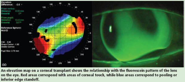
To solve the shortage of the donor cornea in Japan, scientists are developing a poly(vinyl alcohol) hydrogel based keratoprosthesis. Minimum requirements for a keratoprosthesis include light transparency, non-toxicity, and nutrition and fluid permeability. Earlier clinical trials had frequently failed because corneal epithelial down growth occurred between the host cornea and the materials, and the materials were finally rejected from the host cornea. The major cause of this rejection is the weak adhesion between the host cornea and the prosthesis. In order to achieve the firm fixation of the artificial cornea to host cornea, composites of collagen-immobilized poly(vinyl alcohol) hydrogel with hydroxyapatite(PVA-HAp nano composites) were synthesized. The preparation method, characterization, and the results of corneal cell adhesion and proliferation on the composite materials were studied. The PVA-HAp nano composites were successfully synthesized. Chick embryonic keratocyto-like cells were well attached and proliferated on the PVA-Hap composites. This material showed potential for keratoprosthesis.

Introduction
A wide variety of corneal disorders caused by corneal disease and some accidents such as corneal ulcer, chemical burn, etc. are treated by transplantation such as penetrating keratoplasty, lamellar keratoplasty, and deep lamellar keratoplasty, and the success rate is high compared to other tissue transplants. However, many countries suffer from a shortage of donor corneas, and the development of an artificial cornea may be a solution towards solving this problem. In the past 50 years, several groups have attempted to develop reliable artificial cornea, but the trials have frequently failed because of poor biocompatibility. In general, clinically available synthetic devices do not support an intact epithelium, which poses a risk of microbial infection or protrusion of the prosthesis. To solve the problem, we have been developing a poly(vinyl alcohol)(PVA) hydrogel based keratoprosthesis [2-5]. Previously we have found that the immobilization of Type I collagen on the poly(vinyl alcohol)(PVA) hydrogel disc was effective in supporting adhesion and growth of the corneal epithelium and stromal cell in vitro. However, the adhesion was not strong enough to prevent the down growth of the corneal epithelium. Hydroxyapatite(HAp) is well known as one of the best biocompatible materials and it has been applied for percutaneous devices. The result was very promising and the device can prevent epithelial down growth[6-7]. In this study, we synthesized various PVA-hydroxyapatite(PVA-HAp) nanocomposites and modified them by Type I collagen (PVA-COL- HAp) to achieve firm adhesion between the host corneal tissue and the keratoprosthesis. Hydrogel disc of PVA-COL and PVA-COL-HAp and nanofiber sheet of PVA-COL and PVA-COL-HAp were prepared to compare the efficacy of the materials and the influence of the morphology. The preparation method, characterization, and the results of the corneal cell adhesion and proliferation on the composite materials were studied.

By CH . ANKITA 11307010 BME 3rd Year

February 18, 2010

Review on Patents for Mechanical Stimulation of Articular Cartilage Tissue Engineering

Filed under: Uncategorized — bmeforum @ 6:33 am

To repair articular cartilage defects in osteoarthritic patients with three-dimensional tissue engineered chondrocyte grafts, requires the formation of new cartilage with sufficient mechanical properties. The premise is that mechanical stimulation during the culturing process is necessary to reach this aim. Therefore, mechanical stimulation systems have been integrated in aseptic bioreactors for in vitro cultivation of tissue engineered cartilage. These vary from simple unconfined compression systems to advanced bioreactors in which deformation and loading are fully controlled. Fluid handling in these devices is another decisive parameter for the success of cartilage tissue engineering.

Over the last decades bioreactor developments have resulted in the filing of many patents. The aim of this paper is to review these patents, categorize them according to their possibilities for mechanical stimulation and fluid handling systems and finally to discuss them in the context of the demands of a functional tissue engineered cartilage from a mechanical perspective.
Shruti-11307048

OZONE TREATMENT FOR SLIPDISC

Filed under: Uncategorized — bmeforum @ 6:26 am

Absence of Ozone gas– an unstable, allotropic form of Oxygen– in upper reaches of atmosphere means trouble for mankind but its presence in small quantities in spinal cord may do wonders to treat problems relating to disc prolapse. Surgeons have obtained very encouraging results with Ozone therapy in treating disc prolapse cases, commonly known as slip disc.
This is a common lifestyle related problem. Usual causes are faulty posture, obesity, lack of exercise and sudden physical exertion. Though most cases of slip disc can be treated by simple bed rest and and painkillers, some require further treatment or even surgery. It is a very safe procedure and can be carried out in presence of other serious medical problems where general anaesthesia and surgery are not possible. For this reason, it is emerging as safe, relatively painless, and less expensive alternative to surgery. It is an OPD procedure and patient goes home in two or three hours. Until now, no serious side-effects have been observed. It basically involves injecting small amounts of ozone gas generated through a special machine into the affected disc. What ozone does is slowly fragment the disc into its water soluble component parts that get absorbed by the body through normal process.

Ultrasound Imaging of the Musculoskeletal System:

Filed under: Uncategorized — bmeforum @ 6:11 am

Ultrasound imaging, also called ultrasound scanning or sonography, involves exposing part of the body to high-frequency sound waves to produce pictures of the inside of the body. Ultrasound exams do not use ionizing radiation (as used in x-rays). Because ultrasound images are captured in real-time, they can show the structure and movement of the body’s internal organs, as well as blood flowing through blood vessels.
Ultrasound imaging is a noninvasive medical test that helps physicians diagnose and treat medical conditions.

Ultrasound images are typically used to help diagnose:
 tendon tears, such as tears of the rotator cuff in the shoulder or Achilles tendon in the ankle.
 abnormalities of the muscles, such as tears and soft-tissue masses.
 bleeding or other fluid collections within the muscles, bursae and joints.
 small benign and malignant soft tissue tumors.
 early changes of rheumatoid arthritis.

DONE BY
S.SAI SOWMYA(11307038

Localized Hyperthermia Treatment of Cancer.

Filed under: Uncategorized — bmeforum @ 5:35 am

Localized hyperthermia (sustained heating of tissues to temperatures of about 42-43.5 degree C) is one of a number of unconventional methods for treating cancer that are currently receiving increased attention from oncologists. This paper gives a brief review of the effects of hyperthermia on malignant cells and tissues and of methods for producing localized hyperthermia in animals and humans. Radiofrequency and microwave apparatus developed at RCA Laboratories for clinical applications of localized hyperthermia is described in some detail. Clinical results with a variety of cancers are encouraging.

DONE BY
M.SWATHEE(11307052)

February 17, 2010

Auditory Brain Implant.(HArdika Panchani-11307024)

Filed under: Uncategorized — bmeforum @ 8:42 pm

Auditory brainstem implant
An auditory brainstem implant (ABI) is a small device that is surgically implanted in the brain of a deaf person whose auditory nerves are lacking or damaged. The auditory nerves conduct the sound signals from the ear to the brain. The implant enables otherwise deaf people to have a sensation of hearing.

The hearing sensation is limited, but the implant recipients are relieved of total sound isolation, facilitating lip-reading.

The auditory brainstem implant consists of a small electrode applied to the brainstem, a small microphone on the outer ear, and a speech processor. The electrode stimulates vital acoustic nerves by means of electrical signals and the speech processor digitally transmits the sound signals to a decoding chip placed under the skin. A small wire connects the chip to the implanted electrode attached to the brainstem. Depending on the sounds, the electrode delivers different stimuli to the brainstem making deaf people hear a variety of sounds.

Inner ear implants
An inner ear implant, or cochlear implant (CI) is an electronic device surgically implanted in the inner ear of a profoundly or completely deaf individual.

Unlike hearing aids, the cochlear implant does not make sounds louder or clearer. Instead, it stimulates the hearing nerve directly. A cochlear implant gives the hearing impaired recipient a sensation of hearing. It is important to understand that it provides a reduced sense of hearing, only, not a fully restored hearing.
An inner ear implant is comprised of internal and external components. The microphone and speech processor are the external components, with the microphone located on the ear and the processor placed immediately behind the ear. The processor is fixed onto the transmitter implanted beneath the skin.
Another internal component is the decoder placed in the inner ear.

Sounds transmitted directly to inner ear
A cochlear implant takes over the function of the damaged cochlear in the ear, in which some of the vital hair cells are missing. The implant converts speech and surrounding sounds into electrical signals and sends these signals to the hearing nerve in the inner ear. On their way, the signals pass the damaged part of the hearing system. These signals are recognised as sounds by the brain.
Approximately one month after surgery the speech processor is connected and the user will begin to perceive sound. The last part of the implant process is rehabilitation during which the patient receives auditory training and learns different communication techniques. It takes time, practice and patience to learn how to use a cochlea implant.

Chew On This! A Hearing Aid For Your Mouth.

Filed under: Uncategorized — bmeforum @ 8:02 pm

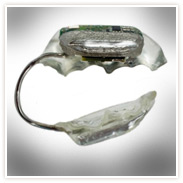
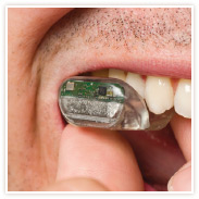
A company in California is developing a hearing aid that will attach to the upper molars and transmit sound through a patient’s jaw. The SoundBite, as it’s called, has two components. The first, a small microphone sitting in the ear canal, records vibrations that it relays to a transmitter resting on back of the ear. These two pieces are attached by a small transparent wire. The second part of the device clamps onto the back of the top row of teeth like an acrylic mold. A receiver detects signals from the transmitter behind the ear and converts them into vibrations strong enough to carry through the teeth and jaw, but weak enough that the wearer can’t consciously feel them.

In a healthy person, air vibrations travel down the inner ear and create patterned disturbances in air pressure that in turn disturb the fluid of the cochlea. Little hair-like cells (called…hair cells) respond to the fluctuations as a spectrum, which the brain then sorts and deciphers as the audible frequencies of sound. The process is one of the most elegant in the body and utilizes the smallest bones in our body. (Take a look!) Tiny, tiny structures convert air vibrations, to fluctuations in air pressure, to wave patterns in fluid, to electrical impulses.

A bone conductive hearing aid, such as the SoundBite, essentially does the same thing, but sends the information through different material. Vibrations ultimately arrive at the cochlea and cause waves patterns in the inner ear, but they get there through surrounding bone (the ear is essentially encased in bone). They are an alternative hearing aid most useful to patients who have ear deformities or infections restricting them from wearing a conventional device.

One of the main problems with bone conductive hearing aids has been keeping them anchored to the bone. The latest approach to doing this has been to surgically implant the hearing aid into the bone surrounding the ear.

SoundBite seems to be an attempt to circumvent the need for surgery, and indeed devises a reversible solution. But there are a lot of other problems that I imagine surfacing when we invite technology into our mouths. Images show that the device hides almost completely in the back of your mouth. So, great. That solves some cosmetic concerns. But what about eating? What about talking? What about keeping it clean? What about talking to, and hearing, your dentist while you’re getting a root canal? Also, people already report poorer sound quality with bone conductive hearing aids than with air conductive ones. Will the quality further deteriorate when the vibrations are coming all the way from the teeth and jaw bone?

By Pratibha  Reg No 11307029 BME 3rd Year

ELECTROPORATION-“KNIFE FOR CANCER”

Filed under: Uncategorized — bmeforum @ 7:47 pm

Electroporation, a technique that microbiologists have long used experimentally to temporarily punch holes in cell membranes and ferry drugs or genes into them, may yield new benefits for cancer treatment, an electroporation device that it claims can kill cancerous tumor cells with remarkable specificity while inflicting little or no damage on surrounding structures and causing no pain for the patient.Electroporation does not produce enough heat to disrupt nearby tissue.
When tumours abut especially large blood vessels, another problem arises for thermal ablation. Radiologists call it the heat sink effect. The flow of blood provides convection to the area, cooling it substantially, and it becomes more difficult to maintain temperatures that thoroughly and consistently ablate the tissue. Angio Dynamics’ device, the Nano Knife, might circumvent this problem all together.
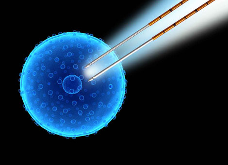
The Nano Knife delivers quick bursts of energy through a set of electrodes inserted into and around the tumour. The pulses can last up to 100 microseconds and create an electrical field of up to 3000 volts per centimetre. A cell within range of the electric field will form pores in its fatty membrane, allowing ions to rush through. When electroporation is performed with a lower voltage than the Nano Knife delivers, and with single pulses instead of a train of pulses, the pores will eventually close as the electric potential of the cell stabilizes. Microbiologists have used this kind of reversible electroporation, among many other things, to transport genetic material into stem cells. When exposed to higher voltages and longer pulse duration, however, the pores in the cell membrane remain open and cause the cell to initiate a programmed suicide, known as apoptosis.
. the Nano Knife has already been approved in the United States for use in the ablation of soft tissue, and Angio Dynamics has installed prototypes in 17 medical centers around the world, 5 of which are actively using it. The device has been tested so far on 37 patients.
Nano Knifes also used to destroy kidney and lung tumours.

Name : AMRITA Reg No : 11307005 3rd Year

Biomedical insights into regeneration of oral tissues

Filed under: Uncategorized — bmeforum @ 5:14 pm

Recent development of biomedical engineering as well as basic biology and medicine has enabled us to induce cell-based regeneration of body tissue assisted with the self-repairing potential tissue or substitute biological functions of damaged organs with cells. For successful tissue regeneration, it is indispensable to give cells an environment suitable for induction of cell-based tissue regeneration. Tissue engineering is a newly emerging biomedical technology to create the environment for tissue regeneration with various biomaterials. Overviews, recent researches and clinical data about oral tissues regeneration based on tissue engineering and the key technologies of tissue engineering come into picture while dealing with such an issue.

Next Page »

Blog at WordPress.com.